Right ovary measures 2.9cm x 2.0cm appears normal. There is no cardiac pulsation seen.
Easy What Is Fetal Cardiac Pulsation For Weight Loss, On transvaginal ultrasound a heart beat can usually be seen at 6 weeks. The general fetal age by weeks can be.
 Apical Pulse Vital Sign Measurement Across the Lifespan 1st From ecampusontario.pressbooks.pub
These shortcuts direct blood away from the lungs and the liver. Tracking a baby’s heart rate helps doctors determine the fetus’s age and growth. The fetal heart rate starts at a slower rate but keeps increasing every day until stabilising at the 12th week. There is well defined single intrautrine gestational sac with good decidual reaction measuring 9.4mm corresponding to 4weeks and 5days.
Apical Pulse Vital Sign Measurement Across the Lifespan 1st Tracking a baby’s heart rate helps doctors determine the fetus’s age and growth.
The transducer is positioned to record through the heart and the changes in response to heart contraction are. Fetal cardiac position refers to the position of the heart within the chest regardless of the fetal cardiac axis or chamber orientation. Embryonic heart pulsation is the earliest proof that the embryo is alive. Hi, i currently in 8 weeks.
 Source: blog.pregistry.com
This movie is a realtime ultrasound recording of the week 12 fetus using doppler to measure the fetal heart rate. By the beginning of the ninth week of pregnancy, the normal fetal heart rate is an average of 175 bpm. The placenta accepts the blood without oxygen from the fetus through blood vessels that leave the fetus. The fetal heart rate starts at a slower rate but keeps increasing every day until stabilising at the 12th week. A Hole in Your Baby’s Heart? The Pulse.
 Source: mdpi.com
At 6 weeks i noticed some clots. On transvaginal ultrasound a heart beat can usually be seen at 6 weeks. Hi, i currently in 8 weeks. Right ovary measures 2.9cm x 2.0cm appears normal. Healthcare Free FullText Heart Rate Assessment during Neonatal.
 Source: researchgate.net
There is no fetal pole. As opposed to levocardia, dextrocardia is a term to describe a heart that is. I did a tvs at 8 week 1 day and they noticed a faint heart beat , the gesational sac size was that of 5 weeks. The fetal heart rate starts at a slower rate but keeps increasing every day until stabilising at the 12th week. Pathophysiology of fetal heart rate changes. Download Scientific Diagram.
 Source: clinicaladvisor.com
By the beginning of the ninth week of pregnancy, the normal fetal heart rate is an average of 175 bpm. To determine how fetal pulse oximetry behaves in various cardiotocographic (ctg) tracings and correlates with neonatal outcome. The placenta accepts the blood without oxygen from the fetus through blood vessels that leave the fetus. The transducer is positioned to record through the heart and the changes in response to heart contraction are. Fetal bradycardia The Clinical Advisor.
 Source: pinterest.com
Embryonic heart pulsation is the earliest proof that the embryo is alive. Fetal heart rate from ultrasound. One side of the placenta is attached to the. The demonstration of early fetal cardiac activity in utero reduces parental anxiety and indicates a favorable prognosis in patients with threatened abortion, and virtually excludes the diagnosis of ectopic pregnancy. Pin by nonas arc on Chest XRay; with the focus on congenital heart.
 Source: obgynkey.com
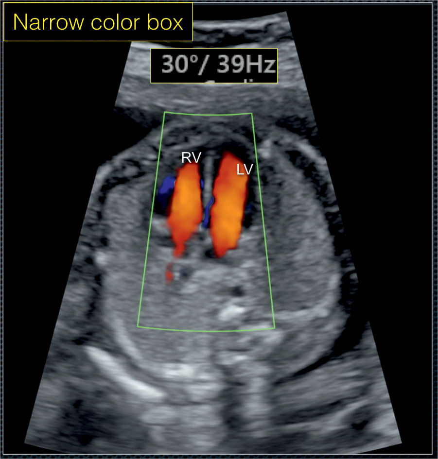
The sections of this tube will go on to form all the structures of the future heart. Embryonic heart pulsation is the earliest proof that the embryo is alive. A fetal heartbeat can be seen and heard during prenatal ultrasound by the sixth week of gestation. Right now the doctor is telling that i am having a. Color Doppler in Fetal Echocardiography Obgyn Key.
 Source: steadyhealth.com
Uterus measures 6.9cm x 5.4cm x 4.1 cm shows a gestational sac. The heart is not fully developed when cardiac activity becomes visible. No scientific evidence backs the claim that the heartbeat varies in boys and girls. This fetal artery connects the pulmonary artery (which will eventually bring blood from the heart to the lungs) and the aorta (which will bring blood from the heart to the body). AFibWhen Heart Patients Do Not Benefit From Aspirin Cardiovascular.
 Source: slideserve.com
A fetal heartbeat can be seen and heard during prenatal ultrasound by the sixth week of gestation. I did a tvs at 8 week 1 day and they noticed a faint heart beat , the gesational sac size was that of 5 weeks. Faint cardiac activity at 8 weeks: At 6 weeks i noticed some clots. PPT Newborn Congenital Heart Disease Screening and Management.
 Source: webstockreview.net
In cases of early pregnancy bleeding, the. Fhr fetal heart</strong> rate is termed a fetal tachycardia and is usually defined as: A fetal heartbeat can be seen and heard during prenatal ultrasound by the sixth week of gestation. To determine how fetal pulse oximetry behaves in various cardiotocographic (ctg) tracings and correlates with neonatal outcome. Heartbeat clipart heart rate, Heartbeat heart rate Transparent FREE for.
 Source: pulse-cardiology.com
At first, all the blood flows into the bottom of the heart, and contractions propel the blood to the top of the tube. Embryonic heart pulsation is the earliest proof that the embryo is alive. What is fetal cardiac arrhythmia? There is also a slowing of the normal fetal heart rate in the last 10 weeks of pregnancy, though the normal fetal heart rate. Congenital Heart Disease, Symptoms, and Treatment Pulse Cardiology.
 Source: holeintheheartasd.org
Tracking a baby’s heart rate helps doctors determine the fetus’s age and growth. Ctg recordings were reassuring or nonreassuring (namely variable or persisting late. Fhr around 170 bpm may be classified as borderline fetal tachycardia. Quick learning videos on radiology for ug and residents in radiologyone of the most important aspect of a fetal anomaly scan is evaluation of the fetal heart. What Is An Echocardiogram? Hole In The Heart.
 Source: blog.pregistry.com
The fetus does not use its own lungs until birth, so its circulatory system is different from that of a newborn baby. In cases of early pregnancy bleeding, the. An arrhythmia is an irregular rhythm of the heart in which abnormal electrical signals through the heart muscle may cause the heart to beat too fast (tachycardia), too slowly (bradycardia), or in an erratic pattern. To determine how fetal pulse oximetry behaves in various cardiotocographic (ctg) tracings and correlates with neonatal outcome. Congenital heart disease ventricular septal defect The Pulse.
 Source: rtmagazine.com
The transducer is initially moved to locate the fetal heart (shown in the upper panel). Tracking a baby’s heart rate helps doctors determine the fetus’s age and growth. The sections of this tube will go on to form all the structures of the future heart. To determine how fetal pulse oximetry behaves in various cardiotocographic (ctg) tracings and correlates with neonatal outcome. Pulse Oximetry for Screening Critical Congenital Heart Defects RT.
 Source: birmingham.ac.uk
As opposed to levocardia, dextrocardia is a term to describe a heart that is. The differential diagnosis of a subtle, t2 bright lesion in the liver includes hemangioma, metastatic disease, and primary liver tumor. By the beginning of the ninth week of pregnancy, the normal fetal heart rate is an average of 175 bpm. Fetal cardiac position refers to the position of the heart within the chest regardless of the fetal cardiac axis or chamber orientation. Call for Europewide screening of babies for heart defects.
 Source: pinterest.com
These shortcuts direct blood away from the lungs and the liver. The differential diagnosis of a subtle, t2 bright lesion in the liver includes hemangioma, metastatic disease, and primary liver tumor. Some cases of fetal arrhythmia are benign, but others can lead to. Quick learning videos on radiology for ug and residents in radiologyone of the most important aspect of a fetal anomaly scan is evaluation of the fetal heart. Pin by nonas arc on SpO2 Congenital heart disease, Pulse oximetry.
 Source: youtube.com
One side of the placenta is attached to the. The average fetal heart rate is between 110 and 160 beats per minute. A slow fetal heart rate is termed fetal bradycardia and is usually defined as 1: It detours blood away from the lungs in utero. बेबी की धड़कन पहली बार कब सुनाई देती है Fetal Heart Rate Chart.
 Source: 99nicu.org
An arrhythmia is an irregular rhythm of the heart in which abnormal electrical signals through the heart muscle may cause the heart to beat too fast (tachycardia), too slowly (bradycardia), or in an erratic pattern. Ultimately, these cells arise from the primitive mesoderm and traverse through the fetal liver to accumulate at the. Sometimes, a heartbeat may not be heard in the early weeks due to inaccurate date calculations, the baby’s position, or the sonography method. Two heart tubes have formed in the embryo. Detecting Congenital Heart Defects After Home Birth All Things.
 Source: med.emory.edu
Fhr fetal heart</strong> rate is termed a fetal tachycardia and is usually defined as: Ctg recordings were reassuring or nonreassuring (namely variable or persisting late. Failure to visualise heart pulsations in embryos measuring 6 mm or less requires patients to wait 7 days for a repeat scan. The transducer is initially moved to locate the fetal heart (shown in the upper panel). Fetal HR Determination Emory School of Medicine.
 Source: obgynkey.com
Hi, i currently in 8 weeks. A slow fetal heart rate is termed fetal bradycardia and is usually defined as 1: Ctg recordings were reassuring or nonreassuring (namely variable or persisting late. Two heart tubes have formed in the embryo. Color Doppler in Fetal Echocardiography Obgyn Key.
 Source: radiologyspirit.blogspot.com
The average fetal heart rate is between 110 and 160 beats per minute. This is because the mother (the placenta) is doing the work that the baby’s lungs will do after birth. To determine how fetal pulse oximetry behaves in various cardiotocographic (ctg) tracings and correlates with neonatal outcome. The heart is not fully developed when cardiac activity becomes visible. RadiologySpirit Early pregnancy (1st trimester).
 Source: blog.pregistry.com
The growing fetus is fully dependent on a special organ called the placenta for nourishment. Fetal cardiac activity (also called fetal heartbeat and usually called embryonic cardiac activity before approximately 10 weeks of gestational age) is the rate of contractions during the cardiac cycles of an embryo or fetus. Embryonic heart pulsation is the earliest proof that the embryo is alive. The heart still has a lot of development to undergo before it is fully formed. What Is Tetralogy Of Fallot? The Pulse.
 Source: verywellfamily.com
This movie is a realtime ultrasound recording of the week 12 fetus using doppler to measure the fetal heart rate. Tracking a baby’s heart rate helps doctors determine the fetus’s age and growth. The growing fetus is fully dependent on a special organ called the placenta for nourishment. The placenta accepts the blood without oxygen from the fetus through blood vessels that leave the fetus. Risk of Miscarriage With Slow Fetal Heartbeat.
 Source: pinterest.com
Tracking a baby’s heart rate helps doctors determine the fetus’s age and growth. There is well defined single intrautrine gestational sac with good decidual reaction measuring 9.4mm corresponding to 4weeks and 5days. Two heart tubes have formed in the embryo. The blood that flows through the fetus is actually more complicated than after the baby is born ( normal heart ). Fetal Circulation Pediatric nursing, Diagnostic medical sonography, Fetal.
 Source: researchgate.net
There is no cardiac pulsation seen. Uterus measures 6.9cm x 5.4cm x 4.1 cm shows a gestational sac. Right ovary measures 2.9cm x 2.0cm appears normal. These shortcuts direct blood away from the lungs and the liver. Pulse wave Doppler during fetal echocardiography showing fetal.
 Source: ecampusontario.pressbooks.pub
There is also a slowing of the normal fetal heart rate in the last 10 weeks of pregnancy, though the normal fetal heart rate. These shortcuts direct blood away from the lungs and the liver. The sections of this tube will go on to form all the structures of the future heart. The heart is not fully developed when cardiac activity becomes visible. Apical Pulse Vital Sign Measurement Across the Lifespan 1st.
I Did A Tvs At 8 Week 1 Day And They Noticed A Faint Heart Beat , The Gesational Sac Size Was That Of 5 Weeks.
There is also a slowing of the normal fetal heart rate in the last 10 weeks of pregnancy, though the normal fetal heart rate. The transducer is initially moved to locate the fetal heart (shown in the upper panel). The sections of this tube will go on to form all the structures of the future heart. At first, all the blood flows into the bottom of the heart, and contractions propel the blood to the top of the tube.
In Cases Of Early Pregnancy Bleeding, The.
Levocardia is a term to describe a heart that is in the normal side of the thoracic cavity (left side) with apex pointing leftward. Cardiac pulsation has been documented in utero by transvaginal scanning as early as 36 days menstrual age, the time when the heart tube starts to beat. The heart is not fully developed when cardiac activity becomes visible. The baby growing inside of the mother’s uterus (the womb) is called a fetus.
The General Fetal Age By Weeks Can Be.
Fhr fetal heart</strong> rate is termed a fetal tachycardia and is usually defined as: The placenta accepts the blood without oxygen from the fetus through blood vessels that leave the fetus. An arrhythmia is an irregular rhythm of the heart in which abnormal electrical signals through the heart muscle may cause the heart to beat too fast (tachycardia), too slowly (bradycardia), or in an erratic pattern. As opposed to levocardia, dextrocardia is a term to describe a heart that is.
There Is No Fetal Pole.
There is no cardiac pulsation seen. These shortcuts direct blood away from the lungs and the liver. Right now the doctor is telling that i am having a. The fetal heart rate starts at a slower rate but keeps increasing every day until stabilising at the 12th week.


